Dr Michael Forrest

Since I first started out in ophthalmology, treatment for glaucoma has focussed on reducing intra-ocular pressure (IOP). Glaucoma, often referred to as the “silent thief of sight,” is a progressive eye disease that damages the optic nerve and can lead to irreversible vision loss. Elevated IOP is the most important risk factor, but many patients with glaucoma have IOP in the “normal” range, and although lowering IOP makes a big difference to most people, and preserves vision for many, it doesn’t restore vision and doesn’t salvage vision for everyone.  The quest for other therapeutic strategies has been an epic, but so far disappointing, enterprise. Interventions that can protect (neuroprotection) or help regenerate (neuroregenration) the delicate retinal ganglion cells (RGCs) and their axons, the retinal nerve fibres that form the optic nerve, remain a Holy Grail. 


Recent advancements have provided a glimmer of hope for patients living with glaucoma.


One such promising avenue of research involves the use of neurotrophic factors – specialized proteins that support the growth and survival of nerve cells in the eye. Ciliary Neurotrophic Factor (CNTF) stimulates the regeneration of RGCs and photoreceptors (the cells in the retina that respond to light and underpin vision). An implant (NT-501) with genetically-modified cells that express CNTF is currently undergoing evaluation in a Phase 2 trial that is due to finish up this year. Recombinant human nerve growth factor (NGF) is also being evaluated, administered as an eye drop. The Phase 1 study of 60 patients was reported in 2022 and showed good tolerability but so far no impact on glaucoma. 


Dietary supplementation with Nicotinamide has shown promise. Nicotinamide is required for the production of NAD+, essential for mitochondria, which act as a cell’s power station. RGCs use a lot of energy, and healthy mitochondrial function is essential. A recent Phase 2 study showed some improvement in visual fields in patients taking nicotinamide and pyruvate supplements.


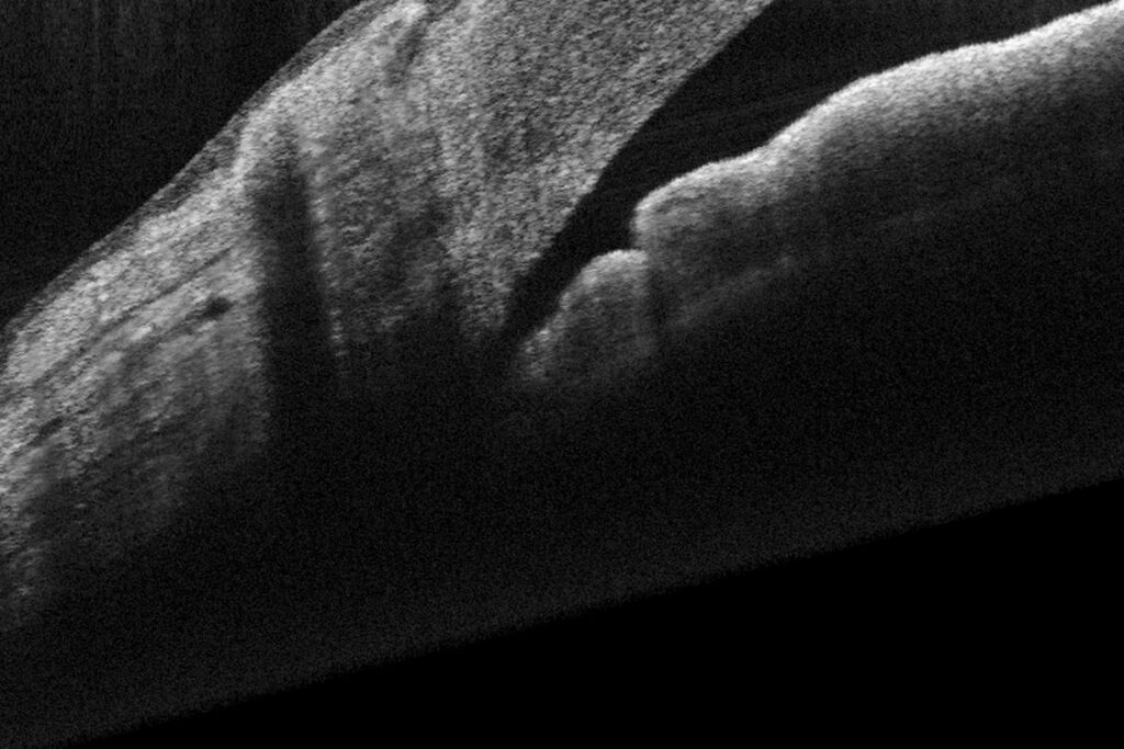
Advances in imaging techniques have allowed for more precise monitoring of the structural changes associated with glaucoma. High-resolution optical coherence tomography (OCT), for example, provides detailed insights into the integrity of the optic nerve and can help us tailor treatment plans to individual patients. Glaucoma management is becoming both more personalised and more proactive.


Challenges remain in the quest for effective support of RGCs in glaucoma, but recent progress in research and clinical practice offer hope for patients facing this sight-threatening disease. By continuing to explore innovative therapeutic approaches and individualized treatment strategies, we move closer to a future with more effective vision preservation.


Beykin G, Stell L, Halim MS, et al. Phase 1b randomized controlled study of short course topical recombinant human nerve growth factor (rhNGF) for neuroenhancement in glaucoma: safety, tolerability, and efficacy measure outcomes. Am J Ophthalmol 2022; 234:223–234.


De Moraes CG, John SWM, Williams PA, et al. Nicotinamide and pyruvate for neuroenhancement in open-angle glaucoma: a phase 2 randomized clinical trial. JAMA Ophthalmol 2022; 140:11–18.

What is "patient-centred care" in cataract surgery? Forrest Eye Care
The variety of IOLs to choose from is enormous

The concept of patient-centred care is not new, but has come to mean a philosophy of heath care. Instead of focussing on a diagnosis, and considering only the treatment of a patient’s disease, patient-centred care means focussing on the individual person, assessing the problems they face, sharing information about options, and collaborating on making decisions about how best to manage those problems. We recognise that each patient is unique, with family experiences, personal preferences, cultural values and traditions, socioeconomic conditions, and lifestyle goals that might be very different from the next person that walks through the clinic door. 

So what does patient-centred care look like in cataract surgery, and what does it mean to me?

Tailoring Treatments to Individual Needs

No two cataract operations are exactly the same.

One key aspect is intraocular lens (IOL) selection.  A synthetic IOL replaces the cloudy, dysfunctional, natural lens removed during surgery. We have choices of monofocal, multifocal and extended depth of focus (EDOF) IOLs (and toric or astigmatism-correcting versions of all of them), so we can address other vision issues like presbyopia and astigmatism, as well as cataract. 

There are advantages and compromises involved with all the available options, and customising care means aligning the treatment with a patient’s lifestyle and visual goals, the condition of their eyes, and allowing a patient to weigh up the relative importance of those various compromises. There is no such thing as a “one size fits all” approach. 

Shared Decision-Making

I think it is essential to have open and honest conversation to understand someone’s  expectations and concerns. A collaborative approach helps us choose a plan that aligns with the patient’s needs.

Asking questions and actively participating in the decision-making process is crucial if a patient is going to achieve the best possible outcome for them, so patients need to be comfortable in the relationship they have with their surgeon.

Preoperative Education and Preparation

Education plays a crucial role in patient-centred care, and part of this is explaining the health of a patient’s eye, and which outcomes are achievable. So the preoperative evaluation assesses more than just the cataract, and needs to be tailored to the individual. For example, the pre-op assessment of someone who had LASIK refractive surgery 15-20 years ago and is otherwise perfectly healthy is likely to be quite different from the assessment of someone with advanced glaucoma, or previous macular surgery. Transparency helps alleviate anxiety and sets realistic goals and expectations.

In the Hospital

Patient-centred care means being concerned with a patient’s experience coming to hospital. The surgical team includes hospital admin staff, nurses, orderlies, anaesthetist, as well as surgeon and surgical assistant, and all of us have a role in alleviating anxiety and making the experience as comfortable as possible.

I’ve always found it important to visit patients in the pre-operative room before surgery, to welcome them and answer any last-minute questions, and I plan my operating lists to allow enough time to do this.  

Follow-Up and Long-Term Care

Postoperative follow-up appointments are vital, because they allow a surgeon to monitor healing and make any necessary adjustments, and because patients often have new questions and concerns that arise. 

People deserve continuous support from their surgeon, and I see every patient I operate on at every post-operative visit. Of course, everyone I operate on has my mobile number in case they need it. 

Improved Patient Satisfaction and Outcomes

Tailoring treatment to individual needs, involving patients in decision-making, and providing comprehensive education and follow-up, means the individual patient’s well-being is at the forefront of every decision. Cataract surgery is about restoring vision and enhancing lives. 

The moment I decided to become an eye surgeon is etched in my memory. I was a medical student at the Princess Alexandra Hospital in Brisbane, watching Professor Lawrie Hirst perform a laser peripheral iridotomy (LPI). The beauty of the eye, the overwhelming importance of preserving vision, and the elegance of the procedure all coalesced in my mind in an instant. I was hooked, and I’ve been hooked ever since. That patient had suffered an attack of primary angle closure glaucoma (PACG), and her second eye was being treated to prevent a similar attack from occurring. 


PACG is not as common as open angle glaucoma, but remains a major cause of irreversible blindness affecting around 20 million people around the world. It occurs in people with “anatomically narrow angles” (ANA), meaning that the front chamber of the eye is shallow enough that in these people the internal flow of fluid becomes obstructed and the pressure inside the eye (the intra-ocular pressure, or IOP) rises catastrophically. PACG can occur at any time, but most commonly occurs at night when the pupil naturally dilates, so treatment is often delayed and results can be compromised. LPI is an essential part of the treatment for patients with PACG, but deciding whether or not to use LPI for prevention in patients with ANA, but without signs of PACG, has been difficult, with unclear evidence to guide decision making. A major randomised controlled trial performed in China, the ZAP (or Zhongshan Angle Closure Prevention) Trial sought to answer the question of whether or not to treat people with ANA, and the 14 year follow-up data have just been published. Also just published this year was a large American study looking at the risk of patients with ANA developing PACG. 


The ZAP Trial showed that although LPI more or less halved the risk of PACG, that risk was fairly low to begin with, and the study authors didn’t recommend using LPI for prevention. It’s very important to note, however, that patients whose IOP rose after they had pupil-dilating drops instilled were treated with LPI and not counted in the study. Many ophthalmologists (me included) would consider that a “provocative test”,  designed to find patients at highest risk. Excluding them from the analysis of results could give a false impression that the risk of PACG is lower than the “real-world” risk. A recent article reviewing the findings of the ZAP Trial (Filippopoulos et al, 2023) suggests that ophthalmologists should still consider LPI for patients with risk factors for PACG (eg hypermetropia, need for frequent dilated retinal exams), or with poor access to medical care. In the real world, I find LPI an important tool to have available. 


A recent case-control study (Yoo et al, 2023) of almost 4000 patients identified through health claims data in the USA reported a rate of conversion from ANA to PACG of around 4% per year. Older age was a risk factor, and for the first time race or ethnicity was not found to be a risk for PACG (although it is probably a risk for ANA with a higher prevalence in Asians). Estimates of rates of risk are hampered by loss to follow-up, and differences in criteria that doctors use to diagnose ANA, but the study is important for showing that cataract surgery is strongly protective against PACG, and the authors recommend considering cataract surgery earlier in patients with ANA and visual symptoms due to cataract. 

References:

Yoo K, Apolo G, Zhou S, Burkemper B, Lung K, Song B, Wong B, Toy B, Camp A, Xu B. Rates and Patterns of Diagnostic Conversion from Anatomical Narrow Angle to Primary Angle-Closure Glaucoma in the United States. Ophthalmol Glaucoma. 2023 Mar-Apr;6(2):169-176. doi: 10.1016/j.ogla.2022.08.016. Epub 2022 Sep 2. PMID: 36058536; PMCID: PMC9978040.


Filippopoulos T, Danias J, Karmiris E, Mégevand GS, Rhee DJ, Gazzard G, Topouzis F, Xu B. Rethinking Prophylactic Laser Peripheral Iridotomy in Primary Angle-Closure Suspects: A Review. Ophthalmol Glaucoma. 2023 Jun 13:S2589-4196(23)00106-0. doi: 10.1016/j.ogla.2023.06.004. Epub ahead of print. PMID: 37321374.


Yuan Y, Wang W, Xiong R, Zhang J, Li C, Yang S, Friedman DS, Foster PJ, He M. Fourteen-Year Outcome of Angle-Closure Prevention with Laser Iridotomy in the Zhongshan Angle-Closure Prevention Study: Extended Follow-up of a Randomized Controlled Trial. Ophthalmology. 2023 Aug;130(8):786-794. doi: 10.1016/j.ophtha.2023.03.024. Epub 2023 Apr 6. PMID: 37030454.

When looking at a near target, such as text on a page (or a phone), the eyes converge so that both are fixating on the object of regard. Convergence Insufficiency, in which the two eyes struggle to maintain fixation together on a near target, is a reasonably common condition in childhood and a cause of double vision, blur at near, and reading difficulty. It has often been treated using vision therapy.

More than 10 years ago the Convergence Insufficiency Treatment Trial (CITT) showed that office-based vision therapy was superior to placebo for treatment of measurable indicators of convergence insufficiency (“near point of convergence” and “positive fusional vergence”). Two follow-up papers from this study were published this week (online pre-publication) in Optometry and Vision Science and show that although these measurable markers of the condition improved with vision therapy, patient reported symptoms (via a patient survey) and, most importantly, reading performance, were not improved by vision therapy any more than by placebo.

The two take-home messages from these trials are:

  1. If you treat convergence insufficiency, use clinical measures, rather than self-reported symptoms, to assess the severity of the condition and the response to treatment; and
  2. if you treat convergence insufficiency, do not claim that it will improve reading skills, because it doesn’t any more than placebo.  

These studies were funded by the National Institutes of Health in the USA, and conducted by academic optometry units across the USA. The study chairperson was Dr Mitchell Scheiman OD PhD, Professor at Pennsylvania College of Optometry and a vision therapist.

References:

CITT-ART Investigator Group . Effect of Vergence/Accommodative Therapy on Reading in Children with Convergence Insufficiency: A Randomized Clinical Trial. Optometry and Vision ScienceOctober 23, 2019.

CITT-ART Investigator Group. Treatment of Symptomatic Convergence Insufficiency in Children Enrolled in the Convergence Insufficiency Treatment Trial–Attention & Reading Trial: A Randomized Clinical Trial. Optometry and Vision ScienceOctober 23, 2019.

This international annual global day of awareness was started in 2000 by Lions Club International, and aims to be a call to action for governments and NGOs, as well as individuals. 

This year the theme is “Vision First”. Worldwide there are more than 250 million people with visual impairment, 36 million of which are blind. More than 75% of these people are suffering avoidable causes of vision loss, and almost 90% of visual impairment is suffered in low and middle income countries.

In Australia, Vision 2020 is focussing on the inequality of health outcomes experienced by people of Aboriginal and Torres Strait Islander descent, and on prevention. We recommend regular eye checks, especially for over-40s, quitting smoking, eye protection for sports and work, regular exercise and following a healthy diet, wearing a hat and sunglasses, and managing diabetes.

#WorldSightDayAU

World Sight Day Forrest Eye Care

Most people having surgery to remove cataracts have been wearing glasses for years by the time their cataracts start to affect vision. Many are well used to glasses by then and ambivalent about wearing them, but an increasing number of people hope to have a degree of independence from glasses. 

Before cataract surgery measurements are taken of a patient’s eyes. This is known as ocular biometry. The length of the eye, curvature of the cornea, depth of the front chamber of the eye and width of the cornea are measured, and several formulae are available to translate these measurements into a useful prediction of which intraocular lens (IOL) to implant to give good unaided distance vision. Lens selection takes quite a lot of time, and happens outside the operating theatre. With modern measurements and modern formulae the results of this prediction are very good, but not perfect. The occasional patient will still require glasses for distance, though these days the vast majority of people maintain driving vision after surgery without glasses.

Improving near vision without glasses is the challenge that lens manufacturers and surgeons are continuing to work on, but at present there are 3 main options for people that wish to improve near vision after cataract surgery: “mono-vision”, multifocal IOLs, and newer “Extended depth of focus” IOLs. All have advantages and drawbacks.

“Mono-vision” means implanting one eye with an IOL for distance and the second eye with an IOL for near. The compromise is that if the second eye is sufficiently myopic to read well, depth perception will usually be lost, and if the second eye is only slightly myopic (“mild” or “mini-“ mono-vision) so that depth perception can be preserved, reading glasses will still be needed for small print. Some people find mono-vision intolerable, although mild mono-vision is much better tolerated.

Multifocal IOLs, which have been around in one form or another for many years now, work by incorporating separate discrete optical zones into the lens. Patients with multifocal IOLs “adapt” to use the individual zones separately, with both bi-focal and trifocal options available.The technologies used to achieve the discrete zones differs between lenses, as does the distance at which near vision is clearest. All multifocal IOLs induce some degree of optical side-effects, usually haloes or “starbursts” around lights at night, presumably due to blur from the other focal zones. These effects can be severe enough that some patients choose to have a second operation to remove the lens and replace it with a monofocal IOL. In addition, because the multifocal IOL splits light between distance, intermediate and near zones (or, in the case of bifocal lenses, distance and near), contrast sensitivity suffers. This makes them inappropriate for most patients with optic nerve disease (including glaucoma) or macular disease (including macular degeneration, epiretinal membrane or diabetic maculopathy).

The latest alternative is the new category of “extended depth of focus” (EDOF) IOLs. These use similar technology to the multifocal IOLs, but induce less myopic defocus so that the risk of both reduced contrast sensitivity and bothersome haloes and glare is much less, and the compromise in distance vision is much less than with multifocal IOLs. The compromise is that these IOLs improve intermediate vision (eg reading your phone), but are not a replacement for reading glasses for most people. 

IOLs that improve unaided near vision have come a long way, and are constantly evolving. For the right patient, well-informed and motivated, they can offer increased freedom from wearing glasses. They are not for everyone, though, which is why they accounted for less than 2.5% of all IOLs implanted world-wide in 2014.

Recently the NSW State Government came under fire for regulating “eyeball tattooing”. In this form of body modification tattoo ink is injected under the conjunctiva to colour the sclera. A tattoo artist, Luna Cobra, claims to have invented the procedure, and to have performed it on around 10 Australians.

This is clearly a specific tattoo niche, with a limited market. But there are major problems trying to regulate novel non-therapeutic procedures, because regulation implies that there is a safe way to perform the procedure. Skin tattooing is an ancient practice, and the earliest known tattoos were found on Otzi, the natural mummy found in South Tyrol, Italy, who lived between 3359 and 3105 BCE. The risks of skin tattooing are fairly well understood. Tattooing the sclera, on the other hand, is new, the long-term hazards are unknown, the risks of the procedure itself are many, and include blindness. The case of a man whose attempted eyeball tattoo resulted in eye penetration and tattoo pigment deposited inside his eye, requiring multiple surgeries to try and repair the damage, was published in August. There seems very little to be gained by attempting to regulate the practice, or in trying to judge who can, and who cannot, perform it safely. The sclera varies in thickness from 1.2mm down to 0.3mm in places, and perforation is a major risk anytime a needle goes near an eye. The needles I use to reattach extraocular muscles during strabismus surgery are specially designed to maintain a level within the sclera and reduce the risk of perforation.

Corneal tattooing has been used therapeutically, to restore a more normal appearance to a blind eye. The most effective way to achieve a good outcome is still not clear.

Kymionis GD1, Ide TGalor AYoo SH. Femtosecond-assisted anterior lamellar corneal staining-tattooing in a blind eye with leukocoria. Cornea. 2009 Feb;28(2):211-3. 

Jalil A, Ivanova T, Bonshek R, Patton N. Unique case of eyeball tattooing leading to ocular penetration and intraocular tattoo pigment deposition. Clin Experiment Ophthalmol. 2015 Aug;43(6):594-6.

There has been a lot of recent discussion in the media about concerns that myopia (short-sightedness) may be increasing among children. Parents are often concerned whether or not they can do something to reduce the chance of their children becoming myopic, or reduce their myopic progression if they already are myopic.


Risk factors for myopia in children include having myopic parents (even after controlling for environmental and demographic differences), and higher socio-economic status. A lot of work has focussed on the impact of near work and time spent indoors. Studies looking at the influence of these factors need to be very carefully designed, because of the potential for confounding bias. People who spend a lot of time outdoors may also be more likely to perform more physical exercise, which needs to be controlled for in study design in case exercise itself is a factor, and may be less likely to spend as much time performing near tasks, so this needs to be controlled for as well.


A number of recent studies have shown that the risk of myopia reduces with increasing time spent outdoors, and increasing light exposure. It also increases with reduced Vitamin D levels, but it is too early to know whether Vitamin D itself plays a role, or whether it is a marker for some other currently unrecognized consequence of light exposure. An interesting recent study in chickens found that intermittent bright light exposure had a greater effect on reducing myopia risk than continuous bright light exposure.


There is much work yet to be done, but so far it seems that putting down the games console and spending a few hours a day running around outside or climbing a tree may be beneficial in ways we parents might not have considered.

Papers:

Lan WFeldkaemper MSchaeffel F Intermittent episodes of bright light suppress myopia in the chicken more than continuous bright light. PLoS One. 2014 Oct 31;9(10).

McKnight CMSherwin JCYazar SForward HTan AX3, Hewitt AWPennell CEMcAllister ILYoung TLCoroneo MTMackey DA. Myopia in young adults is inversely related to an objective marker of ocular sun exposure: the Western Australian Raine cohort study. Am J Ophthalmol. 2014 Nov;158(5):1079-85.

Yazar SHewitt AWBlack LJMcKnight CMMountain JASherwin JCOddy WHCoroneo MTLucas RMMackey DA . Myopia is associated with lower vitamin D status in young adults. Invest Ophthalmol Vis Sci. 2014 Jun 26;55(7):4552-9. 

Guggenheim JAWilliams CNorthstone KHowe LDTilling KSt Pourcain BMcMahon GLawlor DA. Does vitamin D mediate the protective effects of time outdoors on myopia? Findings from a prospective birth cohort. Invest Ophthalmol Vis Sci. 2014 Nov 18;55(12):8550-8.

Karouta CAshby RS. Correlation between light levels and the development of deprivation myopia. Invest Ophthalmol Vis Sci. 2014 Dec 9;56(1):299-309. 

Vaccination is not new technology (Edward Jenner developed the smallpox vaccine in 1796), but it is unquestionably amongst the greatest developments of medical science. Before being wiped out in 1979, smallpox is estimated to have killed 300-500 million people worldwide in the first 79 years of the 20th century.

Anti-vaccinationists are not new either, although, along with climate change denial and skepticism about the moon-landings, vaccine skepticism has grown and continues to take hold of a small but quite vocal group. There are a variety of reasons cited by anti-vaccine campaigners for their stance, including safety concerns, government intrusions, and doubts about efficacy.

But reduced uptake of vaccination has consequences for society as a whole, and especially for those vulnerable patients who are unable to be vaccinated because of immuno-compromise or  other medical conditions. “Herd immunity” refers to the indirect protection enjoyed by unvaccinated people if the vaccinated population is sufficiently large that a disease doesn’t have enough eligible hosts to be spread. The percentage of people in a population that need to be vaccinated to achieve herd immunity is known as the “herd immunity threshold”  (HIT). The HIT is different for different diseases, being relatively low to avoid  influenza pandemics (<50%), higher for smallpox and diptheria (~85%), and much higher (~95%) for measles and pertussis (whooping cough, which kills 1/50 infected infants). Since vaccine uptake in Australia is hovering around 90%, we remain at risk of measles and pertussis outbreaks.

The recent history of Measles in Australia, and the US is very informative. In 2012 the largest measles outbreak in Australia in 15 years occurred, with 173 cases reported, mainly in WA and Southwest Sydney. The initial patient had “imported” the disease from Thailand, but it then easily spread amongst unvaccinated or incompletely vaccinated people. In the first 3 months of 2015 almost 160 people in the US contracted measles, and  three quarters of these were linked to the so-called “Disneyland measles outbreak.” In most otherwise well patients measles is a self-limiting disease. But it can have severe complications. In developing countries corneal complications of measles are an important cause of blindness, with 43 of 193 blind children in a West African cohort blinded by measles. The most feared complication of measles infection is probably subacute sclerosing panencephalitis (SSPE), which causes sight-threatening problems in one third to one half of patients, and ultimately kills 50-90%.

Yesterday the Federal Government announced a policy to tie receipt of government benefits to compliance with the vaccination schedule, and the Federal Opposition joined the government, making this a bipartisan approach. Australians have long benefited from a well-funded vaccination program, with a world-class vaccination register, and it is great to see both sides of politics anxious to increase vaccine uptake. There is certainly some merit in this policy, but we need to remember that people that are scared of vaccination need information and advice, not just direction. The best place to start is with the family doctor.

Marfan Syndrome is a genetic condition that affects connective tissues, particularly the heart valves, aorta, and the eyes. People with Marfan Syndrome also tend to be tall, with long fingers and toes.

In the eye weakness and loss of the zonular ligaments that support the lens lead to lens dislocation (ectopia lentis) and reduced vision. Patients with Marfan Syndrome are also commonly highly myopic (although hypermetropia or far-sightedness can also occur), and have a substantially increased risk of retinal detachment, chronic glaucoma and early cataract. When the lens dislocates so much that glasses or contact lenses can no longer improve vision, surgery is an option. There are some controversies regarding surgical techniques and options for refractive rehabilitation after surgery, but two papers published this year describe significantly different rates of retinal detachment after surgery for Marfan, with a Chinese group reporting 11 Retinal Detachments in surgery on 64 eyes.

Marfan Syndrome was first described by Antoine Marfan, a French paediatrician, at the end of the 19th century. We now know it is caused by mutations in a gene called FBN1, which encodes fibrillin-1, a glycoprotein essential for the elastic fibres in connective tissues. The exact mechanism is not yet clear, but it looks as though fibrillin-1 prevents the accumulation of transforming growth factor beta (TGF-β), and it is likely that reduced fibrillin-1 activity in people with the FBN-1 mutation leads to accumulation of TGF-β, and that this in turn leads to the connective tissue changes of Marfan Syndrome.

When I trained in ophthalmology a combination of systemic signs was required for the diagnosis of Marfan to be made. Someone with ectopia lentis but no other problems was considered to have “isolated ectopia lentis”. We know that eye changes can occur before, and without, significant heart defects in people with Marfan, and few years ago the diagnostic criteria (the so-called Ghent Nosology) was changed so that people with ectopia lentis and either a family history of Marfan, or a FBN-1 mutation, can be diagnosed with the condition. This is important both for assessing their individual risk of developing complications, and for genetic counselling.

In Marfan Syndrome, early diagnosis can reduce the impact of the condition on vision, and reduce the likelihood of cardiac complications. 

Articles

F Fan et  al. Risk factors for postoperative complications in lensectomy-vitrectomy with or without intraocular lens placement in ectopia lentis associated with Marfan syndrome. Br J Ophthalmol. 2014 Oct;98(10):1338-42. 

Miraldi Utz et al. Surgical management of lens subluxation in Marfan syndrome.J AAPOS. 2014 Apr;18(2):140-6.